Home » Posts filed under Obstetrics
 |
| Click at the picture to enlarge |
Introduction:
- A partograph is a graphical record of the observations made of a women in labour
- For progress of labour and salient conditions of the mother and fetus
- It was developed and extensively tested by WHO
- Friedman’s partogram devised in 1954 was based on observations of cervical dilatation and fetal station against time elapsed in hours from onset of labour. The time onset of labour was based on the patient’s subjective perception of her contractility. Plotting cervical dilatation against time yielded the typical sigmoid or ‘S’ shaped curve station against time gave rise to hyperbolic curve.
- The partograph can be used by health workers with adequate training in midwifery who are able to:
o Observe and conduct normal labour and delivery
o Perform vaginal examination in labour and assess cervical dilatation accurately
o Plot cervical dilatation accurately on a graph against time
- There is no place for partograph in deliveries at home conducted by attendants other than those trained in midwifery
- Whether used in health centers or in hospitals, the partograph must be accompanied by a partogram of training in its use and by appropriate supervision and follow up
Objectives:
- Early detection of abnormal progress of a labour
- Prevention of prolonged labour
- Recognize cephalopelvic disproportion long before obstructed labour
- Assist in early decision on transfer, augmentation or termination of labour
- Increase the quality and regularity of all observations of mother and fetus
- Early recognition of maternal or fetal problems
- The partograph can be highly effective in reducing complications from prolonged labour for the mother (postpartum hemorrhage, sepsis, uterine rupture and its sequelae) and for the newborn (death, anoxia, infections, etc)
Functions:
- The partograph is designed for use in all maternity settings, but has a different level of function at different levels of health care
- In health center, the partograph’s critical function is to give early warning if labour is likely to be prolonged and to indicate that the woman should be transferred to hospital (ALERT LINE Function)
- In hospital settings, moving to the right of alert line serves as warning for extra vigilance, but the action line is the critical point at which specific management decisions must be made
- Other observations on the progress of labour are also recorded on the partograph and are essential features in management of labour
Components:
The partograph can be divided into three parts:
- Part 1: Fetal Condition (top)
- Part 2: Progress of labour (middle)
- Part 3: Maternal Condition (bottom)
Outcomes:
[A] Part 1: Fetal Condition
This part of the graph is used to monitor and assess fetal condition:
- Fetal heart rate
- Membranes and liquor
- Moulding of the fetal skull bones
Fetal heart rate | Membrane & Liquor | Moulding of fetal skull bones |
|
(1) Basal fetal heart rate >160 bpm = tachycardia <120 bpm = bradycardia <100 bpm = severe bradycardia | I | Intact Membranes
| 0 | Separated bones, suture felt easily | |
C | Rupture membranes + clear liquor | + | Bones just touching each other | |
M | Rupture membranes + meconium-stained liquor | ++ | Overlapping bones (reducible) | |
(2) Deceleration Yes or No | |
B | Rupture membranes + blood-stained liquor | +++ | Severely overlapping bones (non-reducible) | |
(3) Relation to contraction Early Variable Late | |
A | Rupture membranes + absent of liquor | ↑ molding with head high in pelvis is an ominous sign of cephalopelvic disproportion | |
[B] Part 2: Progress of Labour
This section of the graph has its central feature; a graph of cervical dilatation against time, use to assess the following:
- Cervical dilatation
- Descent of fetal head
- Fetal position
- Uterine contractions
It is divided into a latent phase and active phase:
Latent phase: | Active phase: |
It starts from onset of labour until the cervix reaches 3 cm dilatation | Once 3 cm dilatation reached, labour enters the active phase |
Lasts 8 hours or less | The cervix should dilate at a rate of 1 cm/h or faster |
Contractions at least 2/10 min | Contractions at least 3/10 min |
Each lasting < 20 seconds | Each lasting <40 seconds |
Alert Line (health facility line)
- The alert line drawn from 3 cm dilatation represents the rate of dilatation of 1 cm/hour
- Moving to the right or the alert line means referral to hospital for extra vigilance
Action Line (hospital facility line)
- The action line drawn 4 hours to the right of the alert line and parallel to it
- This is critical line at which specific management decisions must be made at the hospital
[C] Part 3: Maternal Condition
- Name / DOB / Gestation
- Medical / Obstetrical issues
- Assess maternal condition regularly by monitoring:
o Drugs, IV fluids 7 oxytocin, if labour is augmented
o Pulse, blood pressure
o Temperature
o Urine volume, analysis for protein & acetone

a) Body of uterus -
Anatomy |
Position | Pelvic organ at 1st trimester, abdominal organ after 12weeks & dextrorotated as it enlarges due to presence of left side rectosigmoid colon. |
Shape | Pear shape at 1st few weeks, globular shape in the 1st trimester & ovoid shape at term. |
Size | Similar to fetal head at 12th weeks, at the level of umbilicus at 24th weeks & at the level of xiphi-sternum at 36th weeks. |
Consistency | Softening of the uterus is due to ↑ vascularity & presence of amniotic fluid. |
Weight | ↑ from 50-70 gm to 1000 gm at full term (20x ↑ in weight). |
Capacity | ↑ from 10 ml to 5L or more (500-1000x ↑ in capacity) |
|
| Differentiated into deciduas |
| - Hypertrophy & hyperplasia of the muscle fibers due to ↑ synthesis of polyamines. - Differentiated into 3 layers: outer longitudinal- push fetus during delivery, middle interlacing- preventing post-partum hemorrhage & inner circular- hold the fetus up. ↑ elastic fibers facilitating uterine distension. |
| Hypertrophied & firmly attached to the upper uterine segment & loosely attached to the lower uterine segment. (important in caesarean section) |
| Hypertrophy of the broad, round, cardinal & uterosacral ligaments. |
| Blood vessels, lymphatics & nerves. |
|
| Uterine contraction felt during vaginal examination in early pregnancy [these are irregular & painless] |
| Uterine contraction felt during 2nd & 3rd trimester of pregnancy [sporadic, unpredictable, non-rhythmic & its intensity not exceeding 25mm Hg] |
| Uterine discomfort & account for false labour pain as pregnancy advances [↑ in frequency & intensity, no effects on dilation & effacement of cervix] |
Uteroplacental blood flow | Progressive ↑ in uteroplacental blood flow to reach 450-650ml / minute at term |
b) Isthmus - is transformed into the lower uterine segment.
 |
| Upper uterine changes during pregnancy (click at the image to enlarge) |
 |
| Lower uterine changes during pregnancy (click at the image to enlarge) |
- Hypertrophy & softening of the cervix
- Hypertrophy of cervical mucosa
- Increased vascularity of the cervix
- Cervical mucous plug closes the cervical canal due to excess cervical secretion, it is discharged as bloody show with the onset of labor
- Rearragement of collagen fibers
- Hypertrophy & softening
- Varicose veins of the vulva (Kluge's sign)
- Blue or violet discoloration of the vulva (Chadwick's sign)
- Vaginal pH is acidic (3.5 - 6) due to increased production of lactic acid by lactobacilli
- Vaginal cytology show increased small intermediate cells under the effect of progesterone
__________________________________________________________________________________
- Genital System Changes
- Breast Changes
- Cardiovascular Changes
- Respiratory Changes
- Gastrointestinal Changes
- Liver & Gall Bladder Changes
- Urinary Changes
- Skin Changes
- Skeletal Changes
- Nervous System Changes
- Metabolic Changes
- Endocrinal Changes
- Immune System Changes
The items of menstrual history include the following items in sequence:
- [1] Menarche- it is the 1st menses in the woman's life. Normal age is between 9-16.
Precocious puberty: earlier than 9 years old
Delayed puberty: later than 16 years old
- [2] Cycle rhythm- whether the menses occur in regular or irregular period of time.
- [3] Cycle length- it is the duration from 1st day to the next 1st day of menses cycle. Normal cycle length ranges from 21 to 35 days.
<21 days - polymenorrhea
>35 days - oligomenorrhea
- [4] Duration of menstrual flow- It is the period of time during which the menstrual blood flow through the vagina. Normal duration of menstrual flow ranges from 2-7 days.
Menorrhagia: prolonged >7 days and/or 80cc occurring at regular intervals = hypermenorrhoea
Metrorrhagia: irregular uterine bleeding not related to menstruation
Menometrorrhagia: prolonged uterine bleeding occurring at irregular intervals
Polymenorrhagia: frequent heavy/ prolonged menstruation
Postmenopausal bleeding: irregular vaginal bleeding following menopausal cessation of menstruation by 1 year
Intermenstrual bleeding: irregular vaginal bleeding in between regular normal menstruation = breakthrough bleeding
Contact bleeding: irregular vaginal bleeding following tissue contact (examination/intercourse)
- [5] Character of flow- it is regarding the amount, colour and odour of the blood. Normal amount is not more than 80 ml of the total volume. Normal colour of mense blood is dark red.
Excessive flow- indicated by present of clots
Bright red- may indicate excessive flow especially if associated with clots
Polymenorrhea: frequent menstruation occurring at regular intervals (<21 days)
Oligomenorrhoea: infrequent menstruation varying between 35 days and 6 months
Hypomenorrhoea: scanty regular menstruation (<20 ml)
Amenorrhoea: absence of menses for 3 successive regular cycles or 6 months
- [6] Dysmenorrhea- It is pain related to menses. It is severe enough to prevent the woman from doing her daily activities. Dysmenorrhea may be primary or secondary. The tolerable pain at menses is called 'menstrual molimina'.
Primary (spasmodic) dysmenorrhoea
Secondary (congestive) dysmenorrhoea
Special types of dysmenorrhoea
- [7] Intermenstrual Period (IMP)- It is the period from the last day of flow to the next 1st day of flow. Abnormal pain, bleeding or discharge may be presence or absence.
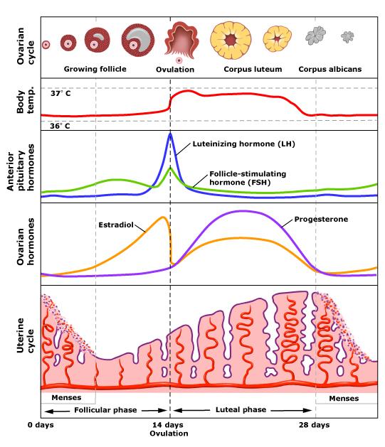
Normally ovulation may cause pain (Mittleschmerz's pain), bleeding (ovulation spotting) or increase of vaginal discharge (ovulation cascade).
- [8] Last Normal Menstrual Period (LNMP)- The 1st day of the last normal menstrual period should be asked for. To certain that the last menstrual period was normal it should fulfill the following criteria:
- It should be normal characters
- It should be preceded by 3 consecutive normal cycles
- It should not be preceded by the use of hormonal contraceptive
NB- The expected date of delivery (EDD) is calculated & mentioned before.
- [9] Current use of contraceptive- It should be mentioned here if it is currently used. The type of contraceptive method and the duration of its use should be asked.
There are 6 procedures in obstetrics maneuver. Each of them has its aim, technique and result. All of these procedures should be performed in sequence because each one depends on the findings of the proceeding procedures.
The 6 obstetric maneuvers are:
The maneuver | The aim |
Fundal level | To determine the uterine size in gestational weeks. |
Fundal grip | To determine which fetal part occupies the fundus uteri. |
Umbilical grip | To determine the position of fetal back. |
First pelvic grip | To determine which fetal part occupy the lower uterine segment. |
Second pelvic grip | To determine the fetal head attitude; whether flexed, deflexed or extended. |
Combined grip | To determine the fetal lie, presentation and fetal tone in case of doubt. |
The ultimate goal of obstetric case taking is to reach the obstetric diagnosis. The items of obstetric diagnosis includes the following in sequence:
Gravidity is the number of pregnancy. It includes the current pregnancy and all other conditions; abnormal pregnancies such as molar and ectopic pregnacies regardless of the outcome; whether delivery or abortion.
Parity is the number of delivery. It includes all of the termination of pregnancies beyond 20 gestational weeks regardless of the fetal outcome; whether living or dead, single or multiple.
- Duration of current pregnancy
It is calculated in weeks using different methods: Naegele's formula and other clinical methods.
It is the relationship between the longitudinal axis of the fetus to the longitudinal axis of its mother.
There are 3 types:
[1] Longitudinal lie [2] Transverse lie [3] Oblique lie
It is the part of the fetus that presents to the pelvic inlet. It is first felt during vaginal examination (PV). There are 3 basics presentation of the fetus which are cephalic (vertex) in 96% of cases, breech in 3.5% of cases and shoulder in 0.5% of cases.
There are 2 theories to explain the high incidence of vertex presentation by adaptation and gravitational theory. In adaptation theory, the fetus more adapted to pyriform-shaped uterus with the larger buttock in the wider fundus and the smaller head in the narrow lower part of the uterus.
The fetal presentation may be determine by denominator.
Denominator is the bony landmark of the presenting part. There are 4 main bone parts to determine the type of fetal presentation which are; chin (face), occiput (vertex), sacrum (breech) and scapula (shoulder).
It is the relation of the fetal back to the anterior abdominal wall of the mother. The fetal back may be antriorly/posteriorly/to the right/to the left of the mother.
The 4 standard positions are left anterior (LA), right anterior (RA), right posterior (RP) or left posterior (LP) in sequence. The occiput (O), mentum/chin (M) and sacrum (S) which are the denominator is important to determine the fetal back position in relation to the mother. All of these bones is directly in relation to the back of the fetus except the chin.

Occipito-anterior positions are more common than occipito-posterior positions because in occipito-anterior positions the concavity of the anterior aspect of the fetus due to flexion fits with the convexcity of the vertebral column of the mother due to its lumbar lordosis.
In each presentation, except the shoulder, there are 8 positions. In vertex presentation they are:
- Left occipito-anterior (LOA) 60%
- Right occipito-anterior (ROA) 20%
- Right occipito-posterior (ROP) 15%
- Left occipito-posterior (LOP) 5%
- Left occipito-transverse (LOT)
- Right occipito-transverse (ROT)
- Direct occipito-anterior (DOA/OA)
- Direct occipito-posterior (DOP/OP)
It means passage of the widest transverse diameter of the presenting part through the plane of the pelvic inlet. The widest transverse diameter of the fetal head is the bi-parietal diameter. The distance between both parietal eminances = 9.5 cm. It is the transverse engaging diameter in all cephalic presentation.
However each cephalic presentation has its own longitudinal engaging diameter according to the attitude of the fetal head (Fetal habitus).
Fetal Habitus is the relation of fetal parts to each other. In majority of cases, the fetus is in the generalized flexion. Some other presentation is extension in face of the fetus.
Synclitism is the 2 parietal bones at the same level. The sagital suture of the fetus is in the midway from symphysis pubis to the promontary of sacrum.
[1] synclitism [2] Posterior asynclitism [3] Anterior asynclitism
Asynclitism is lateral inclination to the frontal head, 2 parietal bones are not at the same level. It is divided into anterior and posterior asynclitism. Anterior asynclitism is more favorable because of less resistance only need to pass promontary of sacrum, less pressure on lower uterine segment and more in axis to the pelvic inlet.
- Complication of current pregnancy
For example; pre-eclampsia, ante-partum haemorrhage, premature rupture of fetal membranes, fetal malformation, fetal death and Rh-iso-immunization.
- Previous disease or cesarean section
For example; maternal cardiac disease, diabetes mellitus, uterine anomalies or fibroids, and previous uterine scar.
So, from the points above, the example of obstetrics diagnosis may be:
"4th gravida, 2nd para, pregnant 36 weeks, longitudinal lie, cephalic presentation, left anterior position, non egaged head, rheumatic heart disease, previous cesarean section."
Contents
- History Taking- (Personal History, Chief Complaint, Menstrual history, Obstetric history, Past History, Family History, Present History)
- Clinical Examination- (General examination, Abdominal Examination, Obstetric Maneuvers )
- Investigation
- Diagnosis
- Fertilization, Implantation, & Early Development
- Plancenta & It's Function
- Amniotic Fluid
- Fetal Circulation
- Maternal Physiological Changes During Pregnancy- (Genital System, Breast, Cardiovascular System, Respiratory System, Gastrointestinal System, Liver & Gall bladder, Urinary System,)
- Diagnosis of Pregnancy
- Vomiting During Pregnancy
- Urinary Tract Disorders With Pregnancy
- Bleeding In Early pregnancy
- Female Pelvis & Fetal Skull
- Normal Labour
- Episiotomy
- Obstetric Analgesia & Anaesthesia
- Abnormal Presentation
- Multiple Pregnancy
- Normal & Abnormal Uterine Action
- Dystocia & Obstructed Labour
- Uterine Rupture
- Genital Tract Injury During Labour
- Third Stage Complications




.png)